Home > Popular Themes > Human Body
Calf muscles, artwork C013 / 4575

![]()
Wall Art and Photo Gifts from Science Photo Library
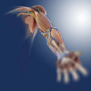
Calf muscles, artwork C013 / 4575
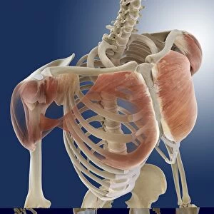
Calf muscles. Computer artwork of a side view of a set of calf muscles (red) and their attachments to the bones of the leg and foot. These are the three fibularis muscles (fibularis longus, fibularis brevis, fibularis tertius) that originate on and run down the fibula, passing through the ankle and attaching to the metatarsals of the foot. The fibularis longus is larger and longer than the other two muscles, which start lower down. These three muscles are used to flex (bend) the ankle downwards and sideways. This view of the left leg includes the knee cartilage, as well as some of the ligaments (white) of the knee, ankle, and foot joints
Science Photo Library features Science and Medical images including photos and illustrations
Media ID 9196475
© SPRINGER MEDIZIN/SCIENCE PHOTO LIBRARY
Ankle Ankles Arthrology Bones Calcaneus Calf Calves Connective Tissue Feet Fibula Foot Heel Bone Joint Joints Knee Knees Lateral Legs Ligament Ligaments Lower Leg Muscles Muscular System Musculoskeletal System Profile Shinbone Side Tendon Tendons Tibia Tibial Musculature
EDITORS COMMENTS
This print showcases the intricate beauty of calf muscles and their attachments to the bones of the leg and foot. In this computer artwork, a side view perspective reveals the three fibularis muscles - fibularis longus, fibularis brevis, and fibularis tertius - originating from the fibula and running down through the ankle before attaching to the metatarsals of the foot. The larger and longer fibularis longus muscle stands out among its counterparts which start lower down. These powerful muscles play a crucial role in flexing (bending) both downwards and sideways at the ankle joint. The detailed illustration also includes other essential components such as knee cartilage, ligaments of various joints including knee, ankle, and foot joints. The image provides an invaluable insight into human anatomy while highlighting key elements like connective tissue, tendons, musculature, ligaments that make up our musculoskeletal system. It is a testament to our body's incredible design for movement. With its focus on biological accuracy and anatomical precision, this print serves as an educational tool for students studying biology or anyone interested in exploring human physiology. It captures not only the functional aspects but also celebrates the aesthetic appeal found within our bodies' structures.
MADE IN THE USA
Safe Shipping with 30 Day Money Back Guarantee
FREE PERSONALISATION*
We are proud to offer a range of customisation features including Personalised Captions, Color Filters and Picture Zoom Tools
SECURE PAYMENTS
We happily accept a wide range of payment options so you can pay for the things you need in the way that is most convenient for you
* Options may vary by product and licensing agreement. Zoomed Pictures can be adjusted in the Cart.