Home > Europe > Italy > Lazio > Rome
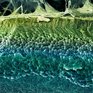
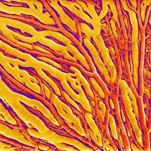
False-colour SEM of retina featuring central fovea

![]()
Wall Art and Photo Gifts from Science Photo Library
False-colour SEM of retina featuring central fovea
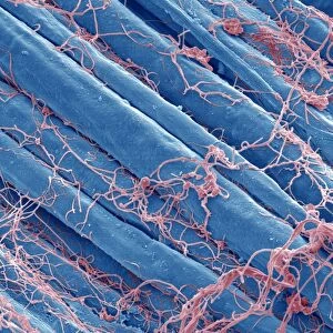
Foveal retina. False-colour scanning electron micrograph of the human retina featuring the central fovea, a crater-like depression in the photosensitive layer of the eye. The foveal retina is the area of greatest visual acuity and contains only cone receptor cells. When an eye looks at an object, that part focused on the fovea is the portion most accurately registered by the brain. This area is particularly rich in blood vessels, which appear root-like, running in, over & outside of the foveal " ridge". Magnification: x24 at 6x7cm size
Science Photo Library features Science and Medical images including photos and illustrations
Media ID 6422336
© PROF. P. MOTTA/DEPT. OF ANATOMY/UNIVERSITY LA SAPIENZA , ROME/SCIENCE PHOTO LIBRARY
Macula Magnified Image Microscopic Photos Retina Sight Subjects Vision Visual Sense False Coloured
FEATURES IN THESE COLLECTIONS
> Europe
> Italy
> Lazio
> Rome
EDITORS COMMENTS
This print showcases a false-colour scanning electron micrograph of the human retina, specifically highlighting the central fovea. The foveal retina is an intriguing crater-like depression found in the photosensitive layer of our eyes. It is within this area that we experience our greatest visual acuity, as it exclusively contains cone receptor cells. When we focus our gaze on an object, the part captured by the fovea is meticulously registered by our brain. This region plays a crucial role in providing us with sharp and detailed vision. As we delve into this microscopic image, one cannot help but notice the abundance of root-like blood vessels that intricately weave their way through, over, and outside of the foveal ridge. With a magnification level of x24 at 6x7cm size, this photograph offers us a glimpse into the intricate world of sight and vision. It reminds us how remarkable and complex our visual sense truly is. Furthermore, it highlights key anatomical features such as the macula and fovea which are essential for optimal visual acuity. Through scientific exploration and advancements in imaging technology like SEM (scanning electron microscopy), we gain invaluable insights into subjects like anatomy and physiology. This particular image from Science Photo Library serves as a testament to both its educational value and aesthetic appeal - showcasing nature's wonders hidden within ourselves without any commercial intent behind it.
MADE IN THE USA
Safe Shipping with 30 Day Money Back Guarantee
FREE PERSONALISATION*
We are proud to offer a range of customisation features including Personalised Captions, Color Filters and Picture Zoom Tools
SECURE PAYMENTS
We happily accept a wide range of payment options so you can pay for the things you need in the way that is most convenient for you
* Options may vary by product and licensing agreement. Zoomed Pictures can be adjusted in the Cart.