White blood cell antigen presentation C016 / 9057

![]()
Wall Art and Photo Gifts from Science Photo Library
White blood cell antigen presentation C016 / 9057
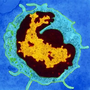
White blood cell antigen presentation. Coloured scanning electron micrograph (SEM) showing the interaction between a macrophage (red) and a T helper lymphocyte (Th cell, yellow), two components of the bodys immune system. Both are types of white blood cell. Macrophages are antigen-presenting cells (APCs). They present antigens (fragments on the surface of pathogens or foreign objects) to T lymphocytes, activating them. Each T lymphocyte recognises and binds to a specific antigen. Binding of the Th cell to the antigen presented by the macrophage activates the Th cell. This leads to its proliferation and the activation of other immune cells that eliminate the antigen. Magnification: x8000 when printed 10cm wide
Science Photo Library features Science and Medical images including photos and illustrations
Media ID 9244415
© STEVE GSCHMEISSNER/SCIENCE PHOTO LIBRARY
Activating Activation Binding Cell Biology Colored Cytological Cytology Electron Microscope Haematological Haematology Hematological Hematology Immune System Immunity Immunological Immunology Interacting Interaction Leucocyte Leucocytes Leukocyte Leukocytes Macrophage Pathogenic Presenting Recognising Recognition T Lymphocyte White Blood Cell Cells Pathogen
EDITORS COMMENTS
This print captures the intricate process of white blood cell antigen presentation within the human body's immune system. The colored scanning electron micrograph showcases a remarkable interaction between a macrophage, depicted in red, and a T helper lymphocyte (Th cell), portrayed in yellow. Both these components play vital roles in defending our bodies against pathogens and foreign objects. Macrophages act as antigen-presenting cells (APCs), presenting antigens found on the surface of harmful substances to T lymphocytes. This interaction activates the Th cell, leading to its proliferation and triggering other immune cells' activation for eliminating the presented antigen. The magnified image, printed at 10cm wide with an impressive x8000 magnification, reveals the minute details of this biological recognition and binding process. Each T lymphocyte possesses unique receptors that recognize specific antigens presented by macrophages. Photographer Steve Gschmeissner from Science Photo Library has masterfully captured this critical moment in cellular biology using a scanning electron microscope. The vibrant colors bring life to this microscopic world, showcasing the complexity and beauty hidden within our immune system's inner workings. This print serves as a testament to both scientific discovery and artistic expression, providing viewers with an awe-inspiring glimpse into one of nature's most fascinating processes – our body's defense against harmful invaders.
MADE IN THE USA
Safe Shipping with 30 Day Money Back Guarantee
FREE PERSONALISATION*
We are proud to offer a range of customisation features including Personalised Captions, Color Filters and Picture Zoom Tools
SECURE PAYMENTS
We happily accept a wide range of payment options so you can pay for the things you need in the way that is most convenient for you
* Options may vary by product and licensing agreement. Zoomed Pictures can be adjusted in the Cart.