Hip anatomy, artwork C013 / 4430

![]()
Wall Art and Photo Gifts from Science Photo Library
Hip anatomy, artwork C013 / 4430
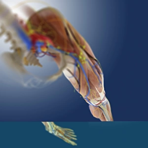
Hip anatomy. Computer artwork of the head of the left femur (centre right) articulating with the left-hand side of the pelvis to form the hip joint. The bones of the left side of the pelvis are semi-transparent here to show details of the head of the femur. The femoral head and pelvic socket include hyaline cartilage (white) and synovial membrane (white) and fluid. Both cartilage and fluid help to lubricate the joint. At the centre of femoral head is its ligament (ligamentum capitis femoris, fibrous white). The pelvic girdle is at lower centre and the coccyx (tailbone of the spine) is at upper left
Science Photo Library features Science and Medical images including photos and illustrations
Media ID 9196009
© SPRINGER MEDIZIN/SCIENCE PHOTO LIBRARY
Arthrology Ball And Socket Joint Bones Capsule Cartilage Coccyx Connective Tissue Femoral Femoral Head Femur Fluid Hip Bone Hips Hyaline Cartilage Ilium Innominate Bone Ischium Joint Joints Legs Ligament Ligaments Lower Back Lower Limbs Musculoskeletal System Osteology Pelvic Pelvic Girdle Pubis Sacrum Skeletal Synovial Membrane Tailbone Thigh Thigh Bone Vertebral Column Articular Cartilage Pelvis
EDITORS COMMENTS
This print showcases the intricate and fascinating anatomy of the hip joint. In this computer artwork, we are presented with a detailed view of the head of the left femur articulating with the left-hand side of the pelvis, forming a vital connection known as the hip joint. The semi-transparent bones on the left side of the pelvis allow us to appreciate every minute detail of this complex structure. The hyaline cartilage, depicted in white, along with synovial membrane and fluid play crucial roles in lubricating and maintaining this joint's functionality. At its core, we can observe the ligamentum capitis femoris, a fibrous white ligament that adds stability to this weight-bearing joint. Surrounding elements such as the pelvic girdle at lower center and coccyx (tailbone) at upper left provide additional context for understanding how these structures relate within our musculoskeletal system. This image not only highlights key components like bones and joints but also emphasizes connective tissues essential for movement and support. It serves as a valuable resource for studying osteology, arthrology, and overall human anatomy. With its remarkable level of detail and scientific accuracy, this artwork from Springer Medizin/Science Photo Library offers an insightful glimpse into one aspect of our body's incredible complexity - reminding us just how intricately designed we truly are.
MADE IN THE USA
Safe Shipping with 30 Day Money Back Guarantee
FREE PERSONALISATION*
We are proud to offer a range of customisation features including Personalised Captions, Color Filters and Picture Zoom Tools
SECURE PAYMENTS
We happily accept a wide range of payment options so you can pay for the things you need in the way that is most convenient for you
* Options may vary by product and licensing agreement. Zoomed Pictures can be adjusted in the Cart.