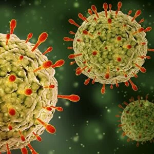
Sindbis virus, computer model

![]()
Wall Art and Photo Gifts from Science Photo Library
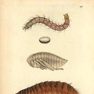
Sindbis virus, computer model
Sindbis virus. Computer model of sindbis virus created using UCSF Chimera molecular modelling software and data from cryo-electron microscopy. It shows the outer glycoprotein shell of the virus. Beneath this layer is a lipid bilayer, an inner protein shell (known as the capsid) and a space containing ribonucleic acid (RNA). Sindbis virus is transmitted by mosquitoes and can cause fever and rash. Cryo-electron microscopy uses beams of electrons, which are fired at multiple angles, to image specimens kept at minus 150 degrees Celsius. The resulting slices of data are reconstructed into 3-D models on computer
Science Photo Library features Science and Medical images including photos and illustrations
Media ID 6412782
© UCSF CHIMERA/SCIENCE PHOTO LIBRARY
3 D Electron Microscopy 3 D Visualisation 3 D Visualization Alphavirus Chimera Computer Graphic Computer Rendering Cryo Electron Cryo Electron Microscope Cryo Em Cryoelectron Microscopy Electron Cryomicroscopy Electron Density Electron Microscopy Glycoprotein Infectious Disease Macromolecular Macromolecule Modelling Molecular Imaging Particle Protein Database Protein Shell Reconstruction Shape Sindbis Virus Structural Biology Surface Ucsf Chimera Viral Viral Shell Virion Virology Viruses Micro Biology Microbiological Molecular Model Molecular Structure Pathogen Protein Virus
EDITORS COMMENTS
This print showcases a computer model of the Sindbis virus, created using UCSF Chimera molecular modelling software and data obtained from cryo-electron microscopy. The image reveals the outer glycoprotein shell of the virus, which is responsible for its infectious nature. Below this protective layer lies a lipid bilayer, an inner protein shell known as the capsid, and a space filled with ribonucleic acid (RNA). Sindbis virus is primarily transmitted by mosquitoes and can lead to symptoms such as fever and rash in infected individuals. To capture this intricate structure, cryo-electron microscopy was employed. This cutting-edge technique involves directing beams of electrons at various angles towards specimens maintained at an extremely low temperature of minus 150 degrees Celsius. The resulting slices of data are then reconstructed into detailed three-dimensional models on a computer. The significance of this visual representation extends beyond its aesthetic appeal; it serves as a valuable tool for researchers in fields like biology and medicine who study viruses and their impact on human health. By providing insights into the structural aspects of pathogens like Sindbis virus, scientists can better understand their behavior and develop effective strategies to combat them. This remarkable photograph exemplifies how advancements in technology enable us to delve deeper into the microscopic world, unraveling mysteries that were once hidden from our sight but now hold immense potential for medical breakthroughs.
MADE IN THE USA
Safe Shipping with 30 Day Money Back Guarantee
FREE PERSONALISATION*
We are proud to offer a range of customisation features including Personalised Captions, Color Filters and Picture Zoom Tools
SECURE PAYMENTS
We happily accept a wide range of payment options so you can pay for the things you need in the way that is most convenient for you
* Options may vary by product and licensing agreement. Zoomed Pictures can be adjusted in the Cart.