Plantarflexion of the foot, artwork C016 / 6800

![]()
Wall Art and Photo Gifts from Science Photo Library
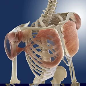
Plantarflexion of the foot, artwork C016 / 6800
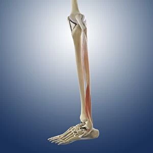
Plantarflexion of the foot. Artwork of the muscles of the foot from the side, with red arrows showing the direction of movement when flexing the foot in the direction of its lower (plantar) surface (plantarflexion). The main muscles involved are the gastrocnemius and soleus (calf muscle), flexor hallucis longus, flexor digitorum longus, and tibialis posterior. These muscles of the lower leg extend downwards into the foot. The nerve used is the tibial nerve. This is the left foot. For the right foot, see C016/6799
Science Photo Library features Science and Medical images including photos and illustrations
Media ID 9245837
© D & L GRAPHICS / SCIENCE PHOTO LIBRARY
Ankle Arthrology Bend Bending Biomechanics Calf Muscle Diagram Flex Flexing Flexion Foot Gastrocnemius Joint Lateral Ligament Ligaments Limb Movement Moving Muscles Muscular Physiological Physiology Plantar Profile Range Of Movements Soleus Tendon Tendons Tibial Nerve Tibialis Posterior Cutouts Extend Extending Flexor Digitorum Longus Left Foot Musculature
EDITORS COMMENTS
This print showcases the intricate artwork C016 / 6800, depicting the plantarflexion of the foot. From a side view, this illustration highlights the muscles of the foot and their movement when flexing towards its lower surface. The red arrows elegantly guide our eyes through this fascinating process. The main players in this graceful dance are revealed to be the gastrocnemius and soleus muscles, commonly known as the calf muscle. Alongside them, we find the flexor hallucis longus, flexor digitorum longus, and tibialis posterior working harmoniously to create this mesmerizing motion. These muscular wonders extend from our lower leg into our precious feet. As we delve deeper into understanding this phenomenon, it becomes apparent that all these movements are orchestrated by none other than the tibial nerve. This particular artwork focuses on capturing these details for our left foot; however, if you seek insight into your right foot's mechanics, refer to C016/6799. With a white background providing contrast against vibrant illustrations, every element is meticulously presented for clarity and comprehension. This print invites us to explore not only anatomy but also physiology and biomechanics with its comprehensive depiction of tendons and ligaments. D & L GRAPHICS / SCIENCE PHOTO LIBRARY has masterfully crafted an artistic representation that seamlessly blends science with aesthetics in this remarkable piece of anatomical artistry.
MADE IN THE USA
Safe Shipping with 30 Day Money Back Guarantee
FREE PERSONALISATION*
We are proud to offer a range of customisation features including Personalised Captions, Color Filters and Picture Zoom Tools
SECURE PAYMENTS
We happily accept a wide range of payment options so you can pay for the things you need in the way that is most convenient for you
* Options may vary by product and licensing agreement. Zoomed Pictures can be adjusted in the Cart.